
Why a Comprehensive Dental Exam Matters
A comprehensive dental exam does far more than check for cavities; it serves as a window into a patient’s overall systemic health. By reviewing medical history, probing periodontal tissues, and evaluating radiographs, dentists can spot early signs of bone‑related diseases such as osteoporosis. Panoramic and intra‑oral X‑rays reveal subtle changes in mandibular cortical width, porosity, and trabecular pattern—radiographic markers that correlate strongly with low bone mineral density in the spine and hip. When thinning of the cortical plate or reduced bone height is observed, especially in post‑menopausal women or older men, the dentist can flag the patient for further evaluation. Integrating risk‑factor questionnaires (age, gender, smoking, calcium intake, menopause) with imaging tools like the Mandibular Cortical Index (MCI) creates a low‑cost, non‑invasive screening protocol that detects skeletal loss before fractures occur. Early identification enables timely referral for a DEXA scan, lifestyle counseling, and preventive treatment, ultimately reducing fracture risk and improving oral outcomes such as implant stability and tooth retention. In this way, a comprehensive dental exam embodies patient‑centered, preventive care that bridges oral and systemic health.
Reading the X‑ray: How Dentists Detect Bone Loss
| Radiographic Modality | Typical Use | Key Indicator(s) for Bone Loss |
|---|---|---|
| Periapical / Bitewing | Interproximal bone assessment | Widened periodontal ligament space, loss of trabecular pattern |
| Panoramic (OPG) | Overall mandibular assessment | Mandibular cortical width (MCW) ≤ 3 mm, eroded cortical index (MCI) |
| Cone‑beam CT (CBCT) | 3‑D bone volume & density | Precise measurement of cortical thickness, trabecular architecture |
| Prior Radiographs | Longitudinal comparison | Year‑to‑year change confirming true bone loss vs. artefact |
All findings are documented per ADA radiographic interpretation standards.
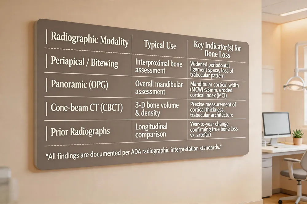 Dentists use routine dental radiographs—periapical, bitewing, panoramic (OPG), and when needed cone‑beam CT—to spot early signs of systemic bone loss. Darker, less radiopaque areas on the alveolar ridge indicate reduced mineral content, while widened periodontal‑ligament spaces and loss of the normal trabecular pattern suggest thinning of the jawbone. Bitewing films are especially useful for interproximal bone loss, whereas panoramic images allow assessment of the mandibular cortical width (MCW) and cortical index (MCI). A MCW of ≤ 3 mm or an eroded MCI (porous or severely eroded cortex) correlates strongly with low bone mineral density and an increased risk of osteoporosis. Dentists compare the current radiograph with prior images to gauge year‑to‑year changes, confirming true bone loss rather than artefacts. All findings are documented according to the American Dental Association’s radiographic interpretation standards. When radiographic clues raise concern, the clinician refers the patient for a dual‑energy X‑ray absorptiometry (DEXA) scan, the definitive test for osteoporosis. Although a dental X‑ray cannot diagnose osteoporosis on its own, it is a valuable, low‑dose screening tool that can trigger timely medical evaluation and preventive treatment.
Oral Clues of Systemic Bone Loss
| Manifestation | Clinical Sign | Underlying Mechanism |
|---|---|---|
| Tooth mobility | Loose or shifting teeth | Thinned alveolar bone reduces support |
| Ill‑fitting dentures | Rapid loss of ridge height/density | Generalized bone resorption of mandibular/maxillary ridge |
| Disproportionate periodontal bone loss | Horizontal bone loss > plaque levels | Systemic demineralization rather than localized periodontitis |
| Delayed socket healing | Slow post‑extraction repair | Compromised bone remodeling capacity |
| Jaw discomfort | Vague pain without obvious pathology | Reduced bone density affecting nociceptive pathways |
Good oral hygiene, adequate calcium/vitamin D, exercise, and smoking cessation can slow progression.
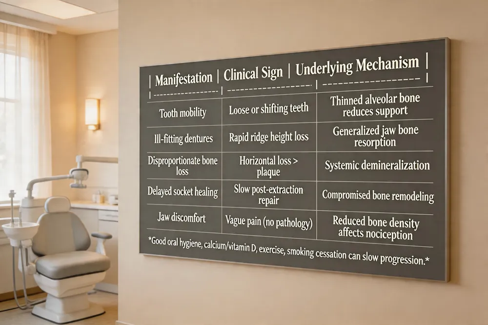 Dentists often encounter early signs of osteoporosis during a routine comprehensive exam. Loose or shifting teeth and an unstable bite are common when the alveolar bone thins, because the supporting jawbone can no longer hold teeth securely. Ill‑fitting dentures appear quickly as the mandibular or maxillary ridge loses height and density, leading to frequent adjustments or replacement. Periodontal bone loss that is disproportionate to plaque levels—especially generalized horizontal bone loss without severe inflammation—suggests systemic bone demineralization rather than localized gum disease. Patients may also report vague jaw pain or delayed healing after extractions, reflecting compromised bone remodeling.
What are the oral manifestations of osteoporosis? The disease reduces jawbone mineral density, visible on radiographs as a thinner, more porous cortical plate and trabecular pattern. This results in accelerated periodontal bone loss, increased tooth mobility, slower socket healing, spontaneous jaw discomfort, and rapid denture‑related bone resorption.
Does osteoporosis cause teeth to break? It does not weaken enamel or dentin directly, but the loss of supporting bone makes teeth and restorations more vulnerable to fracture under normal chewing forces.
Can bone loss in the jaw be reversed naturally? Good oral hygiene, adequate calcium and vitamin D, weight‑bearing exercise, and smoking cessation can slow further loss and modestly promote remodeling, but substantial bone loss usually requires professional interventions such as bone grafts or guided tissue regeneration.
Patient Risk Assessment and Referral Pathway
| Risk Factor | Patient Question (ADA‑style) | Clinical Relevance |
|---|---|---|
| Age & Gender | "What is your age and gender?" | Post‑menopausal women > 65 y have highest osteoporosis prevalence |
| Family History | "Any family history of fractures or osteoporosis?" | Genetic predisposition to low BMD |
| Menopausal Status | "Are you post‑menopausal?" | Estrogen loss accelerates bone loss |
| Calcium/Vitamin D Intake | "How many mg of calcium and IU of vitamin D do you take daily?" | Deficiencies worsen demineralization |
| Smoking & Alcohol | "Do you smoke or consume > 2 alcoholic drinks/day?" | Tobacco & excess alcohol reduce bone formation |
| Physical Activity | "Do you engage in weight‑bearing exercise regularly?" | Sedentary lifestyle contributes to bone loss |
| Radiographic Findings | "Is MCW ≤ 3 mm or MCI eroded on your panoramic?" | Direct imaging evidence of low BMD |
When any of the above are positive, refer for a DEXA scan and initiate lifestyle counseling.
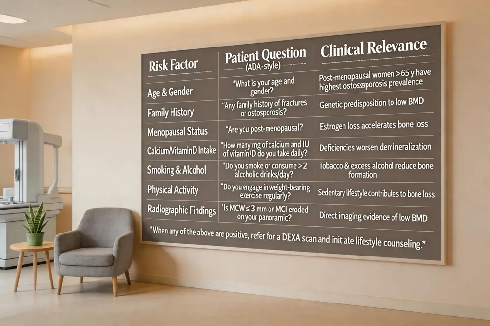 During the medical history review a dentist asks about age, gender, family history of fractures, menopausal status, calcium and vitamin D intake, smoking, alcohol use, and sedentary habits. These key osteoporosis risk factors—genetics, calcium deficiency, tobacco use, menopause, excessive caffeine or alcohol, and low physical activity—are highlighted in ADA‑recommended questionnaires. The first clinical indicator of possible osteoporosis in the dental office is subtle bone loss on panoramic or intra‑oral X‑rays, especially thinning of the mandibular cortical width (≤3 mm) or increased cortical porosity, which often precedes tooth mobility, loose dentures, or ill‑fitting prostheses. When such radiographic changes are observed, the dentist should refer the patient to a primary‑care provider for a dual‑energy X‑ray absorptiometry (DEXA) scan, the gold‑standard test for confirming low bone mineral density. Early detection enables timely lifestyle counseling—adequate calcium (1,200 mg/day for women, 800 mg/day for men, increasing after age 65), vitamin D, weight‑bearing exercise, smoking cessation, and limited caffeine/alcohol—and, if needed, pharmacologic therapy to reduce fracture risk.
Impact on Dental Treatments
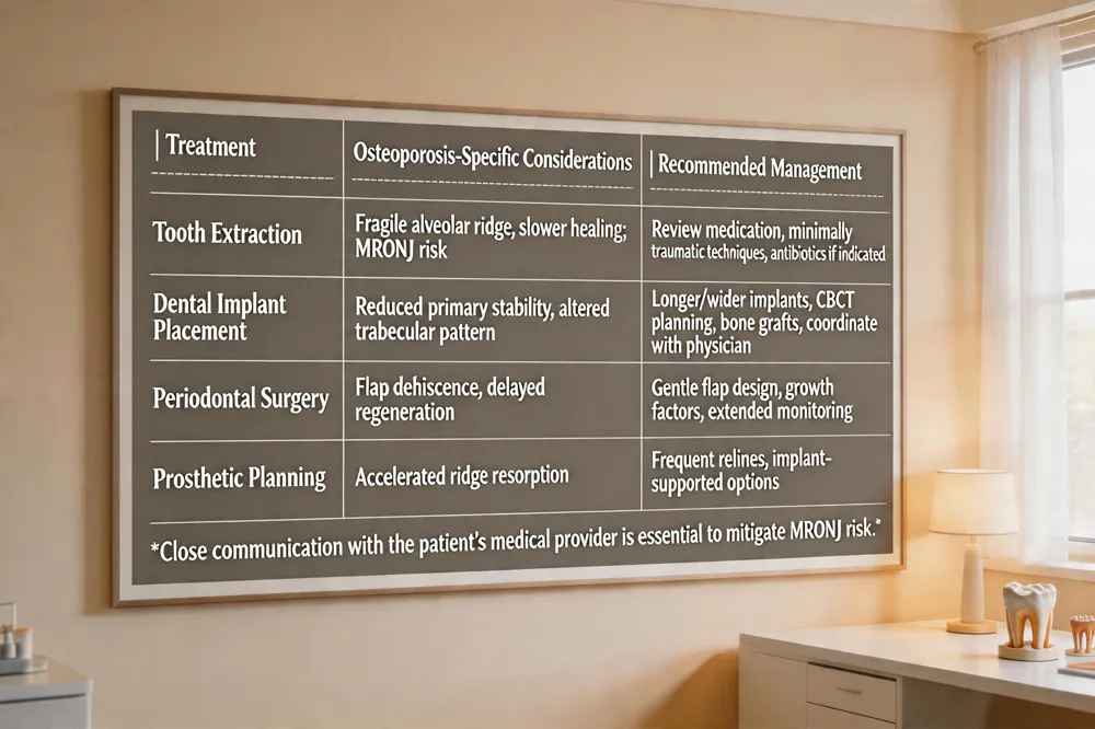
| Treatment | Osteoporosis‑Specific Considerations | Recommended Management |
|---|---|---|
| Tooth Extraction | Fragile alveolar ridge, slower healing; risk of MRONJ with bisphosphonates/denosumab | Review medication, consider drug holiday, use minimally traumatic techniques, prescribe prophylactic antibiotics if indicated |
| Dental Implant Placement | Reduced primary stability, altered trabecular pattern | Use longer/wider implants, perform CBCT planning, consider bone grafts, allow extended osseointegration period, coordinate with physician about anti‑resorptive therapy |
| Periodontal Surgery | Higher risk of flap dehiscence & delayed regeneration | Employ gentle flap design, adjunctive growth factors, extended post‑op monitoring |
| Prosthetic Planning (dentures, bridges) | Accelerated ridge resorption affecting fit | Schedule more frequent relines, consider implant‑supported options when feasible |
Close communication with the patient’s medical provider is essential to mitigate MRONJ risk.
 What considerations are there for dental extractions in patients with osteoporosis?
Patients with osteoporosis have reduced alveolar bone density, making the ridge more fragile and healing slower. Before extraction, the dentist should review the patient’s bone‑health status and any osteoporosis medications—especially bisphosphonates or denosumab—because these increase the risk of medication‑related osteonecrosis of the jaw (MRONJ). Coordination with the prescribing physician about possible drug holidays or prophylactic antibiotics is essential. Gentle, minimally traumatic techniques (periotomes, sectioning, avoiding excessive force) and local hemostatic measures help protect the socket. Post‑operative care includes chlorhexidine rinses, short‑term antibiotics if indicated, and close follow‑up to monitor for infection or delayed bone repair.
What considerations are there for dental extractions in patients with osteoporosis?
Patients with osteoporosis have reduced alveolar bone density, making the ridge more fragile and healing slower. Before extraction, the dentist should review the patient’s bone‑health status and any osteoporosis medications—especially bisphosphonates or denosumab—because these increase the risk of medication‑related osteonecrosis of the jaw (MRONJ). Coordination with the prescribing physician about possible drug holidays or prophylactic antibiotics is essential. Gentle, minimally traumatic techniques (periotomes, sectioning, avoiding excessive force) and local hemostatic measures help protect the socket. Post‑operative care includes chlorhexidine rinses, short‑term antibiotics if indicated, and close follow‑up to monitor for infection or delayed bone repair.
How does osteoporosis impact dental implant placement? Low systemic mineral density and altered trabecular architecture can compromise primary implant stability. Clinicians often compensate by selecting longer or wider implants, employing bone‑augmentation grafts, and allowing a longer healing interval before loading. Cone‑beam CT imaging assesses bone volume and quality, guiding implant design and placement. Communication with the patient’s physician about bisphosphonate or denosumab therapy is crucial to mitigate MRONJ risk. Tailored surgical planning and systemic health monitoring enable safe implant placement even in osteoporotic patients.
How does osteoporosis affect dental treatment planning? Reduced bone quality influences decisions about extractions, implant placement, and periodontal surgery. Dentists may order bone‑density testing, extend healing periods, or use grafting techniques to compensate for weaker bone, ensuring predictable outcomes.
Technology, Costs, and Insurance – Modern Dental Care
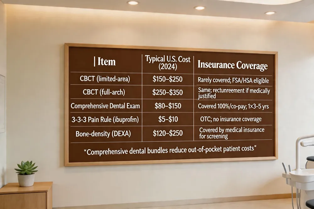
| Item | Typical U.S. Cost (2024) | Insurance Coverage |
|---|---|---|
| CBCT (limited‑area) | $150–$250 per scan | Rarely covered; usually out‑of‑pocket or FSA/HSA eligible |
| CBCT (full‑arch) | $250–$350 per scan | Same as limited‑area; some plans may reimburse if medically justified |
| Comprehensive Dental Exam (incl. full‑mouth X‑rays) | $80–$150 (exam only) | Generally covered 100 % or with modest co‑pay; frequency 1 × 3–5 years per plan |
| 3‑3‑3 Pain Rule (ibuprofen) | $5–$10 for OTC 200 mg tablets | Not an insurance service; over‑the‑counter expense |
| Bone‑density (DEXA) referral | $120–$250 (outside dental office) | Covered by medical insurance when ordered for osteoporosis screening |
Comprehensive dental benefits bundle preventive, restorative, and cosmetic services, reducing out‑of‑pocket costs for patients.
 CBCT imaging benefits
Cone‑beam computed tomography (CBCT) provides three‑dimensional views of the teeth, jawbone, and surrounding structures. It helps dentists assess bone density, plan implant placement, and detect hidden pathology without additional radiation beyond the scan itself.
Pricing of 3‑D scans
In the United States a CBCT scan of the teeth typically costs between $150 and $350 per scan. Prices vary by practice, field‑of‑view size, and whether the scan is bundled with a treatment plan. Larger full‑arch scans tend toward the upper end of the range, while limited‑area scans are nearer the lower end. Insurance rarely covers diagnostic CBCTs, so patients usually pay out‑of‑pocket or use a flexible‑spending account.
Comprehensive dental benefits explained
Comprehensive dental benefits refer to a coverage plan that includes all levels of oral care—from routine prevention to advanced restorative and cosmetic procedures—under a single, coordinated program. They typically pay for regular check‑ups, cleanings, fluoride treatments, sealants, oral‑cancer screenings, digital X‑rays, intra‑oral scans, fillings, crowns, root canals, implants, veneers, orthodontics, and sedation dentistry. By bundling these services, the plan simplifies scheduling and reduces out‑of‑pocket expenses.
Insurance coverage for exams and pain management
Most dental insurance plans cover a comprehensive oral exam (once every 3–5 years) and two routine periodic exams each year, often with full‑mouth X‑rays. Coverage may be 100 % or involve a co‑pay, depending on the policy. The 3‑3‑3 rule—three 200‑mg ibuprofen tablets every three hours for up to three days—is a common over‑the‑counter pain‑management guideline, but severe pain should be evaluated by a professional.
Answer to specific questions
- What do comprehensive dental benefits mean? They provide a “one‑stop” dental home covering preventive, restorative, and cosmetic care, reducing overall costs and improving long‑term oral health.
- How much does a 3‑D scan (CBCT) of the teeth cost in the United States? $150–$350 per scan, with price dependent on field of view and practice location.
- Is a comprehensive dental exam covered by dental insurance in the United States? Yes, most plans include it, often with full‑mouth X‑rays, though coverage details vary.
- What is the 3‑3‑3 rule in dentistry? Take three 200‑mg ibuprofen tablets every three hours for a maximum of three days to manage mild to moderate dental pain.
Empowering Your Smile Through Early Bone Health Detection
A proactive oral‑health monitoring plan turns every routine dental visit into a powerful screening opportunity for systemic bone loss. During a comprehensive exam, the dentist reviews the medical history, probes periodontal tissues, and evaluates panoramic or intra‑oral X‑rays for signs such as thinning mandibular cortex, reduced cortical width (<3 mm), or increased radiolucency. These radiographic clues—often visible a a year‑to‑year comparison—can signal early osteoporosis long before a fracture occurs.
When such findings appear, the dentist collaborates closely with the patient’s primary‑care physician or endocrinologist. A concise referral, including the specific radiographic measurements (e.g., mandibular cortical index or cortical width) and identified risk factors (age > 50, post‑menopausal status, smoking, low calcium intake), guides the physician toward a confirmatory DEXA scan. This interdisciplinary communication ensures that bone‑health management begins promptly, allowing for calcium and vitamin D supplementation, weight‑bearing exercise, smoking cessation, and, when needed, pharmacologic therapy.
The long‑term benefits of early osteoporosis screening through dental exams are substantial. Patients receive earlier medical intervention, which reduces fracture risk, preserves denture fit, and improves implant success rates. Moreover, integrating bone‑health assessment into routine dental care reinforces a patient‑centered, preventive model that supports overall well‑being while safeguarding the smile.