
Why Early Detection Matters
Detecting oral cancer at its earliest stage dramatically improves survival: five‑year survival exceeds 80% for stage I disease, while it falls below 30% once the tumor has spread. A frequent misconception is that oral cancer is always painful or only affects heavy smokers; many early lesions are painless white (leukoplakia) or red (erythroplakia) patches that patients ignore. Routine dental examinations are the most effective defense—dentists regularly inspect the lips, gums, tongue, cheek lining, and neck, and can spot subtle visual or tactile changes before they become symptomatic. Professional screening, complemented by monthly self‑exams, ensures that persistent sores, ulcers, or lumps are evaluated promptly, allowing biopsies and treatment while the disease is still highly curable.
Understanding the First Visual Clues
Early Visual Clues – Quick Reference
| Sign | Description | Typical Location |
|---|---|---|
| Persistent white patch | Leukoplakia – painless, does not fade after weeks | Lips, tongue, inner cheek, floor of mouth |
| Persistent red patch | Erythroplakia – may be velvety, painful | Same as above |
| Non‑healing ulcer | Open >2‑3 weeks, speckled or indurated border | Tongue, palate, floor of mouth |
| Irregular firm lump | Fixed, asymmetrical thickening | Tongue, gums, floor of mouth, neck |
| Pain or numbness | Unexplained, persistent | Any oral site |
 Early visual clues of oral cancer are often painless and easy to miss. The most common signs are persistent white or red patches—leukoplakia (white) and erythroplakia (red)—that do not fade after a few weeks, appearing on the lips, tongue, inner cheek, or floor of the mouth. Non‑healing ulcers that remain open for more than two to three weeks, especially with a speckled or indurated border, are another red flag. Irregular, firm lumps or thickened areas on the tongue, gums, floor of the mouth, or neck—particularly asymmetrical or fixed—suggest early tumor growth.
First signs of mouth cancer pictures
These appear as the described patches, ulcers, or lumps; a velvety red lesion on the ventral tongue or a sharply demarcated ulcer on the palate may also be seen.
How to detect oral cancer early (including at home)
Perform a monthly self‑exam with a bright light and mirror, inspecting all oral surfaces for patches, sores, or texture changes, and palpate the neck and jaw. Any persistent finding warrants a prompt dental or medical appointment.
What are the 7 warning signs of mouth cancer?
- Persistent ulcer or sore >2 weeks
- White/red patches (leukoplakia/erythroplakia)
- Unexplained lumps or thickening
- Persistent pain or numbness
- Difficulty swallowing or chewing
- Hoarseness or sore throat
- Unexplained weight loss or loose teeth.
Risk Factors & Causes You Should Know
Major Risk Factors
| Risk Factor | Why it matters | Typical Relative Risk |
|---|---|---|
| Tobacco use (smoked & smokeless) | Direct carcinogen exposure to oral mucosa | 10‑30× increase |
| Heavy alcohol use | Synergistic with tobacco, irritates mucosa | 5‑15× increase |
| High‑risk HPV (esp. HPV‑16) | Viral oncogenes in oropharynx/tongue | 3‑7× increase |
| Chronic irritation (poor dentures) | Continuous trauma → dysplasia | 2‑4× increase |
| Sun exposure (lips) | UV‑induced DNA damage | 2‑3× increase |
| Low fruit/veg diet | Reduced antioxidants | 1.5‑2× increase |
| Age >40 | Accumulated exposure & DNA damage | Baseline increase |
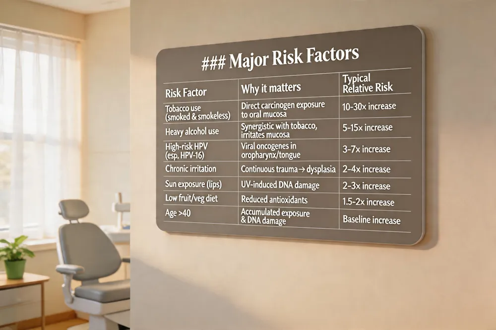 Oral cancer arises primarily from lifestyle and environmental factors that damage the cells lining the mouth. The most significant risk is tobacco use—cigarettes, cigars, pipes, smokeless tobacco—especially when combined with heavy alcohol consumption, which amplifies the danger. Infection with high‑risk human papillomavirus (particularly HPV‑16) has become a major cause, especially for cancers of the tongue and throat. Additional contributors include chronic oral irritation (such as from ill‑fitted dentures), prolonged sun exposure to the lips, a diet low in fruits and vegetables, and genetic predispositions that weaken the immune system. Age also plays a role, with the likelihood of developing oral cancer increasing after the age of 40. Regular dental examinations, self‑exams, and avoidance of tobacco, excessive alcohol, and unprotected sun exposure are key strategies for reducing risk and catching early changes before they become malignant.
Screening, Diagnosis, and the Costs Involved
Screening & Diagnostic Costs (US)
| Procedure | Typical Cost (USD) | Notes |
|---|---|---|
| Routine dental oral (incl. visual exam) | $0‑$150 (often covered) | Part of cleaning visit |
| VELscope fluorescence exam | $19‑$250 | Adjunctive, may be out‑of‑pocket |
| Toluidine‑blue dye test | $0‑$100 | Usually done by clinician |
| Biopsy (incisional/excisional) | $100‑$500 | Often partially covered |
| Imaging (CT/MRI/PET) for staging | $500‑$2,500 | Depends on modality |
| Follow‑up visit | $50‑$200 | Varies by provider |
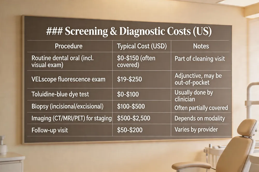 Professional oral cancer exams are a routine part of dental visits; clinicians visually inspect the lips, gums, tongue, cheeks, floor and roof of the mouth and palpate the neck for lumps. Many offices also use adjunctive tools such as VELscope fluorescence or toluidine‑blue dye to highlight suspicious tissue. Patients can complement this with a monthly self‑exam—using a mirror and bright light, look for white (leukoplakia) or red (erythroplakia) patches, non‑healing ulcers, lumps, or changes in texture, and feel the jaw and neck for masses. When a lesion persists, a biopsy (incisional or excisional) is performed, often followed by imaging (CT, MRI, PET) to stage the disease. In the United States, most routine screenings are bundled with a dental cleaning and may be $0‑$150 out‑of‑pocket, depending on insurance. Advanced visual exams (e.g., VELscope) can cost $19‑$250, while a biopsy adds $100‑$500, usually partially covered. No standard blood test exists yet; research into blood biomarkers is ongoing but not yet clinical.
Staging, Pictures, and What Each Stage Looks Like
Staging Overview
| Stage | Tumor Size / Depth | Nodal Involvement | Typical Visual Appearance |
|---|---|---|---|
| 0 (Carcinoma in situ) | Confined to mucosa | None | Flat white/red patch, no ulceration |
| I | ≤2 cm, ≤5 mm deep | N0 | Small painless patch or tiny ulcer |
| II | ≤2 cm >5 mm deep or 2‑4 cm ≤10 mm depth | N0 | Larger patch/ulcer, still localized |
| III | >4 cm or any size >10 mm depth or N1 (≤3 cm node) | N1 | Swelling, firm nodule near jaw/neck |
| IV | Any size with N2‑N3 or M1 | N2‑N3/M1 | Extensive ulceration, necrosis, bone invasion, possible metastasis |
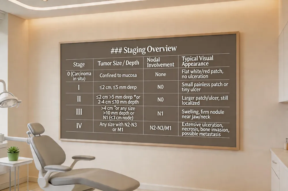 Oral cancer is staged from 0 (carcinoma in situ) to IV using tumor size, depth, lymph‑node involvement, and distant spread. Stage 0 is confined to the mucosal lining with no invasion. Stage I tumors are ≤2 cm and ≤5 mm deep, no nodal disease (T1 N0 M0). Stage II includes lesions ≤2 cm but >5 mm deep or 2–4 cm with ≤10 mm depth. Stage III covers tumors >4 cm, any size with >10 mm depth, or a single ipsilateral node ≤3 cm (N1 M0). Stage IV denotes advanced disease with large or multiple nodes or metastasis (M1). Visually, Stage I often appears as a small, painless white or red patch (leukoplakia/erythroplakia) or a tiny ulcer on the tongue, lip, gum, or cheek. Stage II lesions are larger, deeper, or broader but still localized. Stage III shows swelling or a firm nodule near the jaw or neck, indicating regional node involvement. Stage IV displays extensive ulceration, necrosis, bone invasion, or deformity. High‑quality images for each stage can be found on reputable sites such as the Oral Cancer Foundation, American Cancer Society, and Cancer Research UK.
Treatment Pathways and Prognosis
Treatment Options & Survival Rates
| Modality | When Used | 5‑Year Relative Survival (US) |
|---|---|---|
| Surgery (resection + neck dissection) | Early to locally advanced disease | 80‑86 % for Stage I‑II |
| Radiation therapy (IMRT, proton) | Post‑surgical or definitive for non‑surgical | Improves local control, similar survival to surgery alone |
| Chemoradiation (CT + RT) | Advanced (Stage III‑IV) | 30‑40 % for Stage IV |
| Targeted therapy (cetuximab) | EGFR‑positive tumors, in combo | Modest benefit in select cases |
| Immunotherapy (pembrolizumab) | Recurrent/metastatic disease | Improves survival in PD‑L1 positive tumors |
| Multimodal (surgery + RT ± CT) | High‑risk or bulky tumors | Best outcomes when combined appropriately |
 Mouth (oral) cancer is usually managed by a multidisciplinary team. Surgical removal of the primary tumor and any involved lymph nodes is the cornerstone of treatment; reconstruction of the jaw, palate or soft tissues using grafts and flaps restores speech, swallowing and appearance. When surgery is not feasible or after resection, radiation therapy—often intensity‑modulated, proton or stereotactic—targets residual cells while sparing healthy tissue. Chemotherapy, targeted agents (e.g., cetuximab) and immunotherapy (e.g., pembrolizumab) may be given before surgery to shrink the tumor or combined with radiation (chemoradiation) for advanced disease.
In the United States the overall 5‑year relative survival for oral cavity and oropharyngeal cancer is about 65‑68 %. Localized (stage I‑II) disease enjoys an 80‑86 % survival rate, whereas distant (stage IV) disease falls to 30‑40 %. Early detection dramatically improves these odds; five‑year disease‑free survival for early‑stage cancers reaches 60‑70 %, and many clinicians consider a patient “cured” after five years without recurrence. Regular dental screenings and prompt biopsy of persistent sores, white or red patches, or unexplained pain are essential for achieving the best chance of cure.
Living with Oral Cancer – Symptoms, Reddit Insights, and Reality
Common Patient‑Reported Symptoms (Clinical ↔ Reddit)
| Symptom | Clinical Description | Reddit Mention Frequency |
|---|---|---|
| Persistent white/red patch | Leukoplakia / erythroplakia, painless | High (most posts) |
| Non‑healing ulcer | Open sore >2 weeks, may bleed | High |
| Lump or thickening | Firm, fixed nodule in tongue/neck | Moderate |
| Pain or numbness | Unexplained, sometimes radiates to ear | Low‑moderate |
| Dysphagia / chewing difficulty | Trouble swallowing or chewing foods | Moderate |
| Hoarseness / sore throat | Voice changes without infection | Low |
| Unexplained weight loss | Rapid loss without diet change | Low |
| Loose teeth | Teeth become mobile due to bone involvement | Low |
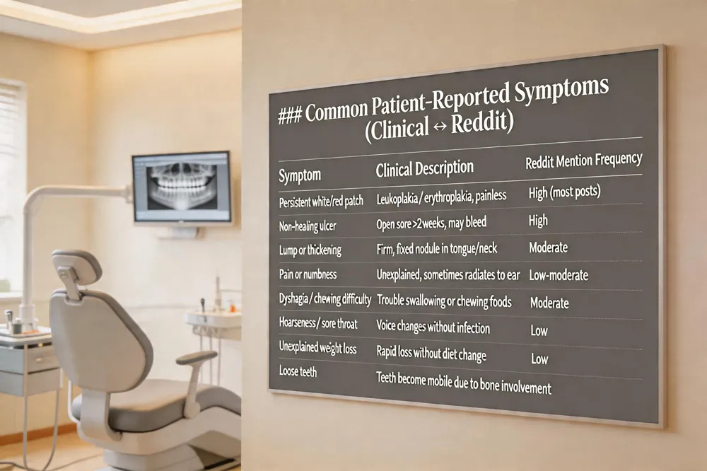 Patients often first notice painless, persistent white (leukoplakia) or red (erythroplakia) patches on the tongue, gums, inner cheek, or lips that refuse to heal for more than two weeks. A non‑healing ulcer, sticky feeling "stuck" in the throat, or a firm lump that feels different from surrounding tissue also signals early disease. Systemic clues such as unexplained weight loss, ear pain without infection, and occasional bleeding while brushing may appear as the tumor grows, even when pain is absent.
Reddit discussions echo these clinical signs: users frequently report lingering sores, color changes, lumps, difficulty swallowing, and a strange sensation in the ear or throat. Many note that the lesions are painless, leading to delayed care.
Can you die from mouth cancer? Yes—advanced-stage oral cancer carries a poor prognosis, with five‑year survival dropping below 30 % for stage IV, while early detection raises survival above 80 %.
Tongue cancer early signs pictures typically show persistent red, white, or dark patches, non‑healing ulcers with raised edges, or a painless nodule on one side of the tongue.
Oral cancer symptoms Reddit: community members commonly mention the above oral changes, ear pain, and weight loss, underscoring the need for prompt professional evaluation.
Take Action Today – Your Smile Depends on It
Regular dental examinations are the cornerstone of early oral cancer detection. During each visit the dentist performs a visual and tactile screening of the lips, gums, tongue, and floor of the mouth, spotting lesions that patients often miss. In addition, a self‑examination at home—using a mirror, bright light, and clean fingers—allows you to notice new white or red patches, non‑healing ulcers, lumps, or changes in texture. If any of these findings persist longer than two weeks, bleeding does not stop, or you experience unexplained numbness or pain, seek professional evaluation promptly. Action dramatically improves treatment outcomes and preserves your smile.