
The Anatomy of Your Smile and Sinuses
Your maxillary sinus cavities are air-filled spaces positioned directly above the roots of your upper molars and premolars. Because these structures share an incredibly thin bony boundary, dental health and sinus comfort are often closely linked. In many individuals, the roots of the upper teeth actually project into the sinus space, creating an environment where dental issues can quickly impact nasal health.
At Empower Your Smile, we emphasize the importance of understanding this anatomical relationship to help you avoid complications. While some practices treat oral and sinus issues as separate concerns, we recognize that up to 40% of maxillary sinus infections have an odontogenic origin. Addressing concerns like persistent pressure or lingering discomfort requires a careful assessment of both your dental anatomy and sinus function.
Our mission centers on patient education, ensuring you have the information needed to distinguish between a routine dental recovery and a potential sinus-related complication. By integrating advanced imaging and comprehensive clinical oversight, we focus on preserving your long-term health, ensuring that your path to a confident smile remains free of unnecessary nasal or sinus distress.
The Anatomy Underlying Your Dental and Sinus Health
 The maxillary sinus cavities are air-filled spaces situated behind your cheekbones, positioned directly above the alveolar process of your maxilla. Because this bone structure also serves as the foundation for your teeth, the roots of your upper molars and premolars often lie in extremely close proximity to the sinus floor. In many cases, these roots project directly into the sinus cavity, creating an intimate anatomical link between your dental environment and your nasal health.
This proximity explains why dental issues and sinus infections frequently produce overlapping symptoms, such as facial pressure or localized pain in the upper jaw. When an infection develops at the root of an upper tooth, it can readily extend into the sinus space, a condition known as odontogenic sinusitis. Research indicates that such dental sources may account for as many as 30% of maxillary sinusitis cases. At Empower Your Smile, we emphasize that distinguishing between a standard toothache and a sinus-related issue is vital for effective treatment.
Diagnostic accuracy is paramount because conventional two-dimensional X-rays often fail to capture the subtle bone changes associated with sinus-related dental pain. In fact, an estimated 86% of odontogenic cases may go undiagnosed by general practitioners who rely only on basic imaging. To bridge this gap, our team at Empower Your Smile utilizes Cone Beam Computed Tomography (CBCT) to gain a precise, three-dimensional view of the sinus-tooth relationship. By identifying potential risks before starting restorative work, we can ensure that every procedure protects your oral health while respecting the delicate anatomy of your sinus floor.
Shared Proximity. The roots of upper teeth are separated from the sinus floor by a thin layer of bone, meaning inflammation can easily travel between these systems.
Shared Symptoms. Pressure or pain in the cheek area is common to both conditions, often intensifying during activities like chewing or when changing head positions.
Diagnostic Precision. Advanced 3D imaging is required to confirm whether a persistent ache originates from a dental infection or a sinus allergy.
Identifying Dental-Origin Sinus Issues
 Dental issues frequently serve as the primary source of maxillary sinus infections. Research indicates that more than 30% of cases involving inflammation in these cavities stem from oral sources, a condition often referred to as sinonasal complications of dental disease or treatment. Because the roots of your upper molars and premolars reside in close proximity to the floor of the maxillary sinus, even separated by a paper-thin layer of bone, bacteria from abscesses or periodontal disease can easily migrate into the sinus.
Common symptoms often include persistent nasal congestion, facial pain, and tenderness in the cheek area. Unlike typical sinus issues, dental-origin discomfort frequently intensifies when biting or chewing. Practitioners at empoweryoursmile.com utilize advanced diagnostic imaging to differentiate these issues from conventional rhinosinusitis, which is vital because up to 86% of dental-related sinus cases may otherwise go undiagnosed by primary care providers per UT Health San Antonio.
Distinguishing between acute and persistent inflammation is critical. Acute symptoms may resolve with standard care, but when dental decay or infection remains, the resulting odontogenic sinusitis often becomes chronic. Relying on antibiotics without addressing the underlying oral cavity pathology—such as an infected tooth or a structural communication—rarely provides long-term relief.
- Can dental issues cause sinus problems? Yes, dental issues can definitely lead to sinus problems. The roots of your upper back teeth are situated very close to—or may even extend into—the maxillary sinus cavities located behind your cheekbones. When these teeth develop infections, decay, or other dental pathologies, that inflammation can easily spread to the sinuses, potentially resulting in chronic or recurrent sinusitis. Furthermore, side effects of dental distress, such as mouth breathing due to persistent tooth pain, can contribute to a dry mouth that exacerbates overall oral health issues. Because symptoms of dental problems and sinus infections often overlap, it is important to consult your dentist to rule out or treat any underlying oral health concerns.
Navigating Complications During and After Procedures
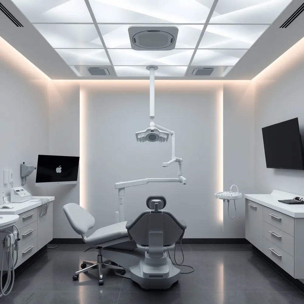 Because the roots of the upper teeth are located in close proximity to the maxillary sinuses, dental procedures can occasionally impact these air-filled spaces. The extraction of upper molars can sometimes create an accidental opening between the mouth and the sinus cavity, known as an oroantral fistula or sinus perforation. Beyond physical openings, existing dental infections or unresolved abscesses can allow bacteria to travel directly into the maxillary sinus, triggering inflammation and infection. Additionally, professional dental work may occasionally aggravate pre-existing sinus sensitivities or shift pressure within the area. At Empower Your Smile, we utilize advanced imaging like CBCT scans to carefully map these anatomical structures, significantly minimizing the risk of complications compared to clinics relying solely on traditional two-dimensional X-rays.
What are the common symptoms of a sinus perforation after a tooth extraction?
Patients should monitor for specific physical indicators if they suspect a communications issue following an extraction. Common symptoms include persistent one-sided nasal congestion, a distinct sensation of air movement between the mouth and nose, or fluid leaking from the oral cavity into the sinus. Other signs of sinus perforation include a whistling sound when breathing, recurring nasal discharge, or a pressure-like sensation in the cheekbone area. Small openings may heal naturally with proper post-operative care, but any persistent discomfort lasting beyond a few days warrants a professional evaluation to ensure the sinus membrane remains intact and protected.
| Procedure Type | Primary Risk | Preventative Focus |
|---|---|---|
| Tooth Extraction | Sinus Perforation | Radiographic Depth Assessment |
| Dental Implants | Membrane Intrusion | Precise CT Planning |
| Root Canal | Localized Inflammation | Bone Proximity Mapping |
Distinguishing Toothache from Sinus Pain
Distinguishing between general dental pain and sinus-related discomfort can be difficult because the roots of your upper molars sit in close anatomical proximity to the maxillary sinus cavities. Standard toothaches typically stem from localized issues like cavities or pulpitis and often present as sharp sensitivity to temperature or specific pressure while biting.
Sinus-related pain, in contrast, frequently manifests as a dull, steady ache affecting multiple upper teeth simultaneously. You may notice this discomfort intensifies during fluctuations in head position, such as when bending forward, or during periods of active nasal congestion. At empoweryoursmile.com, our experts emphasize that while a singular tooth problem is often isolated, sinus-induced pressure typically correlates with broader symptoms like facial fullness, postnasal drip, or a foul-smelling discharge per UT Health San Antonio.
If you experience dental sensitivity alongside persistent sinus blockage or altered smell, the problem may be odontogenic sinusitis. Because conventional X-rays may miss up to 86% of dental-origin sinus cases, seeking a formal diagnostic evaluation is critical. A comprehensive dental exam provides the necessary imaging to accurately identify whether you need treatment for a specific tooth or care for an underlying sinus pathway.
What to Expect During Your Recovery
It is not uncommon to experience mild sinus discomfort following dental procedures, particularly those involving your upper molars. Because the roots of these teeth are located in close proximity to the maxillary sinus floor, dental work can sometimes cause temporary inflammation of the sinus membrane. While this is often a brief, expected side effect, persistent or worsening symptoms should not be ignored.
At Empower Your Smile, we prioritize your long-term oral and nasal health by conducting thorough pre-procedural assessments. If you notice signs such as facial pressure, congestion, or discomfort that does not subside after a few days, it is important to contact our Tribeca office for an evaluation. We are committed to your comfort and will ensure any post-treatment issues are addressed promptly to keep your smile healthy and pain-free.
- Avoid forceful nose blowing for several days after surgery to minimize pressure changes that could disrupt healing.
- Maintain consistent oral hygiene, including recommended saline rinses, to prevent the buildup of bacteria in the surgical site.
- Avoid smoking, as it is a significant risk factor that can delay healing and increase inflammation in the sinus area.
- Recognize warning signs that require professional attention, such as fever over 101°F, pus-like nasal discharge, or severe swelling that persists beyond two weeks.
While mild sinus inflammation typically resolves within one to two weeks, as noted in recent clinical observations, patients should remain vigilant. Persistent symptoms or the emergence of new, sharp pain could indicate sinonasal complications of dental disease or treatment that require intervention through a comprehensive dental exam or other diagnostic imaging.
Prioritizing Your Long-term Oral and Nasal Health
Because the anatomy of your maxillary sinuses is so closely linked to your upper teeth, even minor dental issues can cause persistent sinus discomfort. Research shows that up to 70% of unilateral maxillary sinusitis cases trace back to a dental origin, per Radiopaedia. Relying on conventional imaging often reveals little, as nearly 86% of these conditions go undiagnosed by primary providers, noted by UT Health San Antonio.
At empoweryoursmile.com, we prioritize accuracy by integrating advanced diagnostic technology for every patient. Whether you require routine maintenance or specialized restorative work, our team ensures your oral health is evaluated in the context of your total wellness. By catching hidden cavities or assessing bone density before procedures, we minimize the risk of complications while providing comprehensive dental care. If you experience persistent cheek tenderness or unusual tooth sensitivity, contact our practice today to ensure your smile and sinuses remain in balance.