Why Crown Lengthening Matters for Implants
Crown lengthening is a periodontal surgery performed by a periodontist that removes excess gum tissue and, when needed, a small amount of alveolar bone to expose additional tooth or implant platform structure. By re‑establishing the biologic width (≈2–3 mm) and creating a sufficient vertical dimension, the procedure ensures a stable, healthy gingival margin and adequate bone height for the prosthetic crown. This improves oral hygiene, reduces plaque traps, and prevents chronic inflammation that can compromise osseointegration. Consequently, implants placed after proper crown lengthening achieve better primary stability, more natural emergence profiles, and higher long‑term success rates, often exceeding 95 % when case selection and healing protocols are followed.
Understanding Crown Lengthening: Basics and Surgical Details
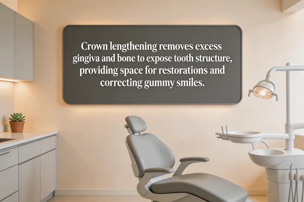 Crown lengthening surgery is a periodontal procedure that removes or repositions excess gingival tissue—and when necessary, a small amount of underlying bone—to expose additional natural tooth structure. It is indicated for subgingival decay, fractures, short clinical crowns, or to correct a gummy smile, providing a stable foundation for crowns, bridges, veneers, or implant restorations. The periodontist numbs the area with local anesthesia (sedation optional) and then creates a flap incision, reshapes tissue and bone, and stabilizes the flap with fine sutures. Typically absorbable sutures (e.g., Vicryl) dissolve within a week, while non‑absorbable silk stitches are removed after 7‑10 days. Patient comfort is maintained by the anesthesia during surgery and mild postoperative soreness managed with ibuprofen or acetaminophen, ice packs, and a soft‑food diet. Most patients resume normal activities within a day, and full soft‑tissue healing occurs within 1‑2 weeks, with complete stabilization up to three months.
Pre‑Operative Planning, Coding, and Implant Guidelines
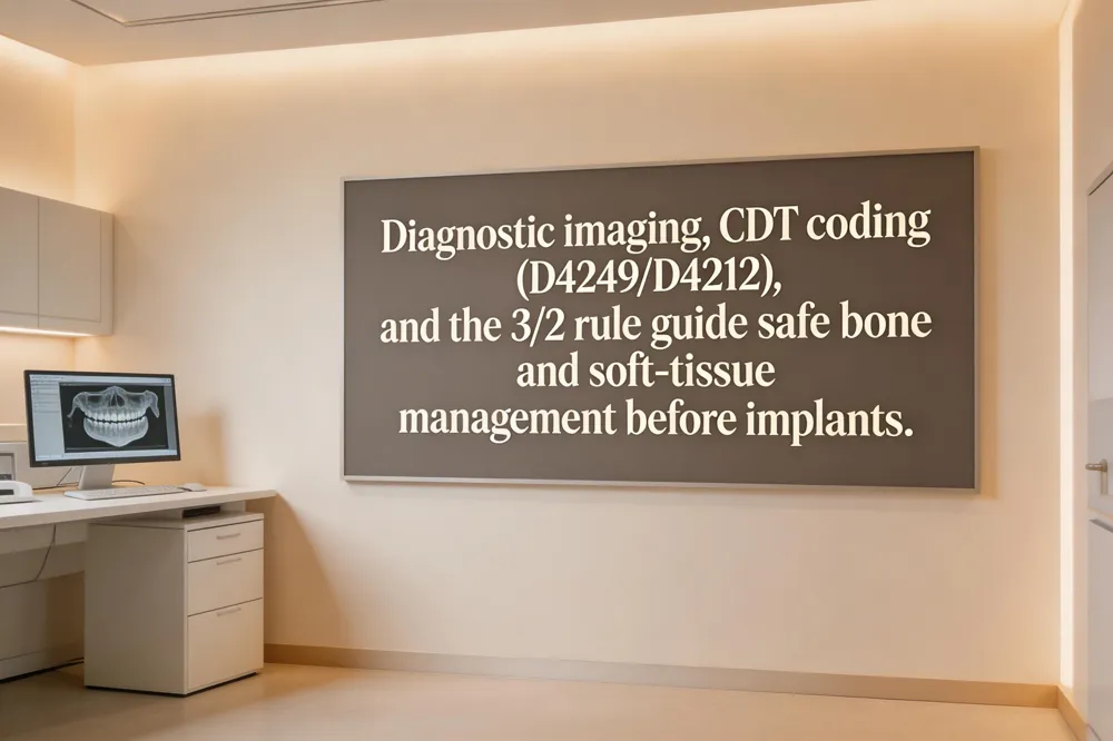 Successful crown‑lengthening before implant placement begins with a thorough diagnostic work‑up. A cone‑beam CT (CBCT) together with intra‑oral scans maps the distance from the cement‑enamel junction to the bone crest, allowing the clinician to verify that at least 3 mm of supracrestal tooth structure will remain after surgery—this preserves the biologic width (≈2 mm) and establishes a stable ferrule.
Coding – The primary CDT code for hard‑tissue crown lengthening is D4249 (Clinical crown lengthening – hard tissue). When only soft‑tissue removal is needed, use D4212 (Gingivectomy or gingivoplasty for restorative access). Traditional CPT/ADA equivalents are D4210 for gingival‑tissue surgery and D4211 when bone removal is performed. Document clinical indications, radiographs, and a six‑week healing interval to avoid claim denials.
Implant Placement Rules – The 3/2 rule mandates a minimum of 3 mm vertical bone height above the implant platform and 2 mm horizontal bone thickness around the implant to ensure long‑term stability. Crown‑height space (CHS) for a fixed prosthesis should be 8–12 mm; if CHS is insufficient, bone grafting or a reduced‑crown design may be required.
Crown lengthening code – D4249 for hard‑tissue removal; D4212 for soft‑tissue only.
What is the 3/2 rule for implant placement? – At least 3 mm vertical bone height and 2 mm horizontal bone thickness around the implant.
Crown height space guidelines – Ideal CHS is 8–12 mm; less may need grafting or design modification.
Step‑by‑Step Clinical Protocol for Crown Lengthening
 Crown lengthening procedure steps
A case begins with a comprehensive clinical assessment and diagnostic imaging (periapical X‑rays or CBCT) to determine the amount of soft‑ and hard‑tissue removal needed while preserving biologic width. After confirming the plan, local anesthesia (and optional sedation) is administered. A crevicular incision is made, the flap reflected, excess gingiva excised, and, if required, a minimal amount of alveolar bone is contoured to expose at least 3 mm of supracrestal tooth structure. The flap is repositioned apically and fine sutures are placed to hold the tissue in a tension‑free manner. Detailed post‑operative instructions—including gentle oral hygiene, a soft‑food diet, ice packs, and scheduled follow‑ups—are provided to promote optimal healing.
Crown lengthening procedure steps
A case begins with a comprehensive clinical assessment and diagnostic imaging (periapical X‑rays or CBCT) to determine the amount of soft‑ and hard‑tissue removal needed while preserving biologic width. After confirming the plan, local anesthesia (and optional sedation) is administered. A crevicular incision is made, the flap reflected, excess gingiva excised, and, if required, a minimal amount of alveolar bone is contoured to expose at least 3 mm of supracrestal tooth structure. The flap is repositioned apically and fine sutures are placed to hold the tissue in a tension‑free manner. Detailed post‑operative instructions—including gentle oral hygiene, a soft‑food diet, ice packs, and scheduled follow‑ups—are provided to promote optimal healing.
Incision for crown lengthening A crevicular (intrasulcular) incision follows the natural gingival sulcus around the target teeth, often scalloped or beveled to preserve papillae and vascular supply. One or two vertical releasing incisions may be added mesial or distal for access. The flap is reflected to expose bone for precise osteotomy while maintaining soft‑tissue stability.
Functional crown lengthening steps After assessment, anesthesia, and incision, a full‑thickness flap is reflected. Selective osseous recontouring removes bone to achieve the desired tooth length, ensuring at least 3 mm of healthy tissue above the bone crest. The flap is repositioned apically and sutured, followed by postoperative care and a temporary restoration until tissue maturation for the final crown.
Digital, Guided, and Laser‑Assisted Techniques
 Modern crown lengthening relies on a CBCT‑intra‑oral scanning workflow that merges 3‑D bone data with soft‑tissue scans to produce a virtual treatment plan. From this plan a CAD‑CAM, 3‑D‑printed surgical guide (often a double‑guide) is fabricated to dictate incision depth, flap elevation, and precise osteoplasty, preserving biologic width while achieving the desired gingival margin.
Guided esthetic crown lengthening uses that digital design to position the free‑gingival margin and osseous crest accurately. The guide directs tissue and bone removal, increasing predictability, shortening operative time, and delivering a harmonious smile that matches the pre‑operative simulation.
Laser‑assisted esthetic crown lengthening can be performed open‑flap (OF) or flapless (FL). Both achieve similar long‑term margin levels, but FL reduces postoperative pain, swelling, and bleeding, shortens surgery by ~15 minutes, and offers a more comfortable recovery, while OF provides direct bone access for extensive osteotomy.
High‑quality 3‑D animations of the procedure are available on MEDtube, YouTube, Shutterstock, and Envato, illustrating each surgical step for patient education.
Healing Timeline, Diet, and Post‑Operative Care
 After crown‑lengthening, patients should avoid alcohol for at least the first 48 hours and preferably up to two weeks, as alcohol can disrupt clotting and increase bleeding. Stick to soft foods (yogurt, scrambled eggs, blended soups) initially; they are recommended for the first 24–48 hours. By day 3–5 you may introduce semi‑solid foods (oatmeal, well‑cooked pasta) and return to regular solid foods after 3–5 days once sutures are removed, swelling subsides, and chewing feels comfortable. Chew on the opposite side and avoid straws, smoking, and vigorous rinsing.
The gum tissue needs 6–8 weeks to stabilize, and many clinicians wait 2–3 months (up to 6 months in the aesthetic zone) before placing a permanent crown. A temporary restoration protects the tooth during healing.
For a printable guide, request the “Crown Lengthening Post‑Op Instructions” PDF from the office; it outlines medication, ice‑pack schedule, oral‑hygiene steps, and diet recommendations.
Managing Tissue Response and Aesthetic Modifications
 Gingival rebound after crown lengthening is a common early healing phenomenon in which the repositioned gum margin migrates coronally, typically within the first few weeks and stabilizing by three months. The extent of rebound depends on periodontal phenotype, age, and surgical technique; flap‑based approaches (e.g., apically positioned flap) generally produce less regrowth than laser or electrosurgical gingivectomy. Meticulous oral hygiene and gentle handling of the surgical site help limit tissue regrowth, and persistent rebound may require a minor re‑contouring before the final restoration.
In the United States, crown‑lengthening costs range from $300‑$500 for a single tooth to $1,000‑$3,000 per tooth when bone removal is needed, with quadrant fees between $500‑$1,500. Insurance may cover functional cases, while purely cosmetic procedures often require out‑of‑pocket payment.
The Kois technique follows a systematic protocol that begins with incisal‑edge and lip‑dynamic analysis, proceeds to a precise external‑bevel gingivectomy, and, if necessary, uses a KB‑1 straight chisel for controlled osseous reduction while preserving biologic width.
To correct excessive gingival display (a “gummy smile”), clinicians employ conventional gingivectomy, flap surgery with osseous reduction, [laser] (https://www.palmbeachperio.com/a-comprehensive-guide-to-crown-lengthening/) ablation, or guided tissue regeneration. Modifications such as temporary block‑out resin or customized gingival stents stabilize the new margin during healing, and the choice of technique is guided by tissue thickness, biotype, and aesthetic goals.
Clinical Decision‑Making: Crown Lengthening vs. Implants
 When to choose Crown lengthening over implants: Crown lengthening is preferred when the tooth is salvageable, the crown‑to‑root ratio remains favorable (typically >1:1), and the patient wishes to retain the natural tooth. It is also ideal for correcting a gummy smile or creating a ferrule for a restoration. Implants are indicated when the tooth is non‑restorable, the crown‑to‑root ratio is poor, or severe periodontal disease makes preservation risky.
Timing of crown lengthening relative to restoration: The procedure should be completed before the final crown is prepared. By exposing at least 3 mm of supracrestal tooth structure and re‑establishing biologic width, the dentist gains sufficient material for a proper ferrule and ensures long‑term crown stability. Healing typically requires 4–6 weeks for soft tissue and up to 3 months for full bone remodeling before the definitive restoration is placed.
Finding a specialist in New York City: If you’re searching for "crown lengthening near me," Empower Your Smile in Tribeca, NYC, offers board‑certified periodontist services. Dr. Moezinia reshapes gum tissue and, when needed, a small amount of bone to expose more natural tooth structure, delivering both functional and aesthetic benefits. The office provides thorough pre‑operative planning, local anesthesia, and a smooth recovery protocol. Schedule a consultation to discuss your case, insurance coverage, and cost estimate.
Before‑and‑After Outcomes and Patient Experience
 Crown lengthening is often requested by patients who have a “gummy” smile, short clinical crowns, or uneven gingival margins that make teeth look diminutive. Before the procedure, excessive gum tissue may obscure three‑quarters of the crown, creating an imbalanced appearance. After the surgery, the periodontist gently recontours the gingiva and, when necessary, removes a thin layer of bone to expose at least 3 mm of supracrestal tooth structure, restoring a harmonious tooth‑to‑gum ratio. The visual result is a longer, brighter tooth display, a symmetrical gum line, and increased confidence without the need for veneers.
Patient comfort is a priority; local anesthesia eliminates pain, while options such as nitrous oxide or oral sedation are offered for anxious individuals. Empower Your Smile in Tribeca tailors each case with advanced digital planning, laser‑assisted soft‑tissue removal, and custom surgical guides to achieve precise, minimally invasive outcomes. Personalized post‑operative instructions—including soft‑food diets, ice packs, and gentle oral hygiene—ensure a smooth recovery and optimal aesthetic results.
Your Next Steps Toward a Healthier, More Confident Smile
Start by scheduling a comprehensive exam with a board‑certified periodontist. During this visit the clinician will review your medical history, take detailed clinical measurements and digital imaging (CBCT or intra‑oral scans) to evaluate gum thickness, bone levels, and crown‑to‑root ratios. With that data the team will discuss a personalized crown‑lengthening plan—choosing the appropriate technique (gingivectomy, apically positioned flap, or laser‑assisted approach) and, when needed, outlining the amount of bone recontouring required to preserve biologic width and create an optimal emergence profile. Once the soft‑tissue and bone have healed (typically 4–12 weeks), you will proceed to the implant phase, where a guided surgery protocol can place the implant precisely for a long‑lasting, aesthetic restoration. This coordinated workflow ensures functional stability, improved oral hygiene, and a confident smile.